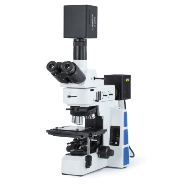

Product Performance


Application Cases
Biomedical field:
It can be used to identify tumor cells, hemorrhagic polyps, sarcoidosis, lymphocytic leukemia, cytoplasmic and nuclear differentiation, and cell counting.
")
Rapid identification of hemorrhagic polyp and fleshy white spot regions of the laryngeal mucosa based on microscopic hyperspectroscopy (red region)

Microscopic hyperspectral discrimination of tumor location and abnormal cell spread under 20x eyepiece
")
")
Rapid differentiation of nuclei, cytoplasm and other material based on microscopic hyperspectral calculations Number of cells based on position of cytoplasmic centers (402 total)
Dark-field scattering nanoparticle detection
Dark-field microscopy is a special microscopic technique realized under dark-field illumination, which prevents light unrelated to the object under observation from entering the objective lens and presents a clear outline of the object against a dark background. Microparticles as small as 4-200 nm can be visualized with a resolution up to 50 times higher than that of normal bright-field illumination microscopy. Microscopes equipped with hyperspectral imaging systems can be used to identify the types of particles.
The figure shows VNIR hyperspectral imaging of lung tissues from mice after a single intratracheal drop of low (18 pg) and high (162 pg) titanium dioxide nanoparticles to determine the location of particle retention in these tissues.
")
Dark-field image of nanotitanium dioxide exposed tissue (top)
Dark-field hyperspectral images from titanium dioxide nanoparticle-exposed tissues identified these nanoparticles, which behaved as aggregates of white inclusions (middle panel)
Titanium dioxide nanoparticles in these tissues show up as red dots or aggregates in hyperspectral maps (below)
OLED display luminescence test
The microscopic hyperspectral imaging system can obtain the light-emitting images of OLED display with higher spatial resolution through different magnification eyepieces, and detect the uniformity and stability of OLED display light-emitting through the feature of "one spectrum in one" of hyperspectral image data.
")
Detection of OLED display luminescence at 20X, 50X and 100X
Wafer material and defect detection
Non-contact, non-destructive, fast and accurate micro-region measurement technology, which can be operated at room temperature or on-line in production, can obtain the PL Mapping of the entire wafer, thus obtaining important information about the group distribution ratio of the substrate or epitaxial layer, defects and other properties of the material in the micro-region of the homogeneity. Based on the micro-hyperspectral imaging technology, the material of the wafer and the change of the concentration of the luminescence center of the sample can be identified on a fine scale.
")
Images and spectra of wafers implanted with boron, aluminum, and non-implanted special materials under microscopic hyperspectral imaging systems
Applications in Chalcogenide Crystals
The micro-hyperspectral imaging system for detecting inhomogeneities in chalcogenides has the following advantages over traditional techniques such as confocal microimaging: single whole-field-of-view imaging; the intensity of the excitation source in the field of view of the system is uniformly distributed in PL imaging experiments; and quantitative values of spectral intensities can be obtained.
")
Chalcogenide PL data. Figures (a) and (b) show two different monochromatic PL images taken at 625 nm and 750 nm, respectively
Figure (c) shows the spectra of different positions in Fig. 1
Figure (d) shows the frequency shift imaging of the PL mapping in the specified region
Applications on LED/OLED light source displays
Microscopic hyperspectral technology is gradually applied in the testing of semiconductor materials and devices. Microscopic hyperspectral imaging technology is mainly used in the study of light-emitting uniformity of semiconductor materials, detection and analysis of defects in semiconductor materials, and spatial distribution of temperature on the surface of LED chips.
")
Micro-hyperspectral inversion of the temperature of panels with different LED light sources
Application Areas
1. Biomedical field
2、Dark field scattering nanoparticle detection
3、OLED display light-emitting test
4、Wafer material
5. Defect detection
6. Application in chalcogenide crystals
7, LED / OLED light source display on the application of